Publications

Recombinant functional multidomain hemoglobin from the gastropod Biomphalaria glabrata.

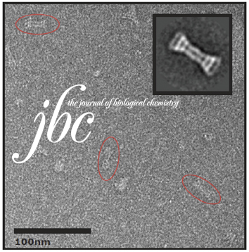
The extracellular hemoglobin multimer of the planorbid snail Biomphalaria glabrata , intermediate host of the human parasite Schistosoma mansoni , is presumed to be a 1.44 MDa complex of six 240 kDa polypeptide subunits, arranged as three disulfide‐bridged dimers. The complete amino acid sequence of two subunit types (BgHb1 and BgHb2), and the partial sequence of a third type (BgHb3) are known.

Recombinant functional multidomain hemoglobin from the gastropod Biomphalaria glabrata. Read More »
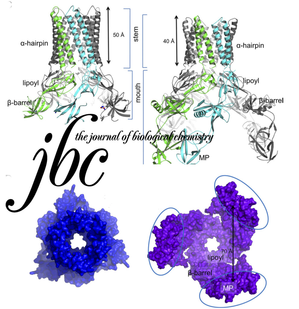
Funnel-like hexameric assembly of the periplasmic adapter protein in the tripartite multidrug efflux pump in gram-negative bacteria.

The Escherichia coli AcrAB-TolC pump is the principal multidrug exporter that confers intrinsic drug tolerance to the bacteria. The inner membrane transporter AcrB requires the outer membrane factor TolC and the periplasmic adapter protein AcrA. However, it remains ambiguous how the three proteins are assembled. In this study, a hexameric model of the adapter protein was generated based on the propensity for trimerization of a dimeric unit, and this model was further validated by presenting its channel-forming property that determines the substrate specificity.

Funnel-like hexameric assembly of the periplasmic adapter protein in the tripartite multidrug efflux pump in gram-negative bacteria. Read More »

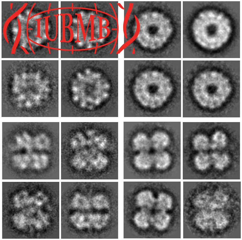
Initial evaluation of a direct detection device detector for single particle cryo-electron microscopy.

We report on initial results of using a new direct detection device (DDD) for single particle reconstruction of vitreous ice embedded specimens. Images were acquired on a Tecnai F20 at 200 keV and a nominal magnification of 29,000×.

Initial evaluation of a direct detection device detector for single particle cryo-electron microscopy. Read More »

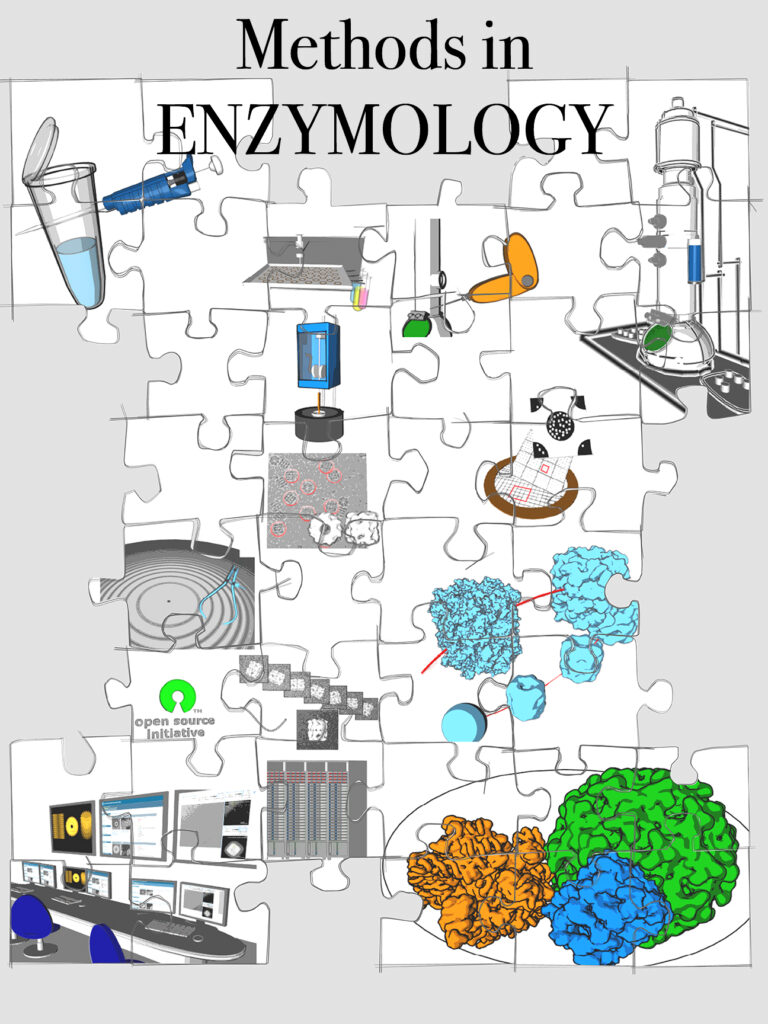
Automation in Single-Particle Electron Microscopy Cryo-EM, Part C: Analyses, Interpretation, and Case studies

Throughout the history of single-particle electron microscopy (EM), automated technologies have seen varying degrees of emphasis and development, usually depending upon the contemporary demands of the field. We are currently faced with increasingly sophisticated devices for specimen preparation, vast increases in the size of collected data sets, comprehensive algorithms for image processing, sophisticated tools for quality assessment, and an influx of interested scientists from outside the field who might lack the skills of experienced microscopists.

Automation in Single-Particle Electron Microscopy Cryo-EM, Part C: Analyses, Interpretation, and Case studies Read More »

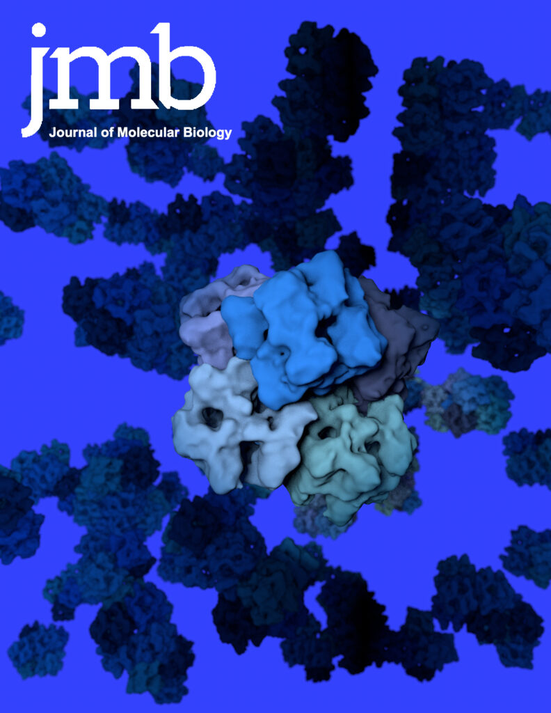
10-A cryoEM structure and molecular model of the Myriapod (Scutigera) 6x6mer hemocyanin: understanding a giant oxygen transport protein.

Oxygen transport in Myriapoda is maintained by a unique 6 × 6mer hemocyanin, that is, 36 subunits arranged as six hexamers (1 × 6mers). In the sluggish diplopod Spirostreptus, the 1 × 6mers seem to operate as almost or fully independent allosteric units (h ∼ 1.3; P50 ∼ 5 torr), whereas in the swift centipede Scutigera, they intensively cooperate allosterically (h ∼ 10; P50 ∼ 50 torr).

10-A cryoEM structure and molecular model of the Myriapod (Scutigera) 6x6mer hemocyanin: understanding a giant oxygen transport protein. Read More »

Comparative 11A structure of two molluscan hemocyanins from 3D cryo-electron microscopy.

Hemocyanins are giant extracellular proteins that transport oxygen in the hemolymph of many molluscs. Molluscan hemocyanins are cylindrical decamers or didecamers of a 350–400 kDa subunit that contains seven or eight different covalently linked globular functional units (FUs), arranged in a linear manner. Each FU carries a single copper active site and reversibly binds one dioxygen molecule. As a consequence, the decamer can carry up to 70 or 80 O2 molecules.

Comparative 11A structure of two molluscan hemocyanins from 3D cryo-electron microscopy. Read More »

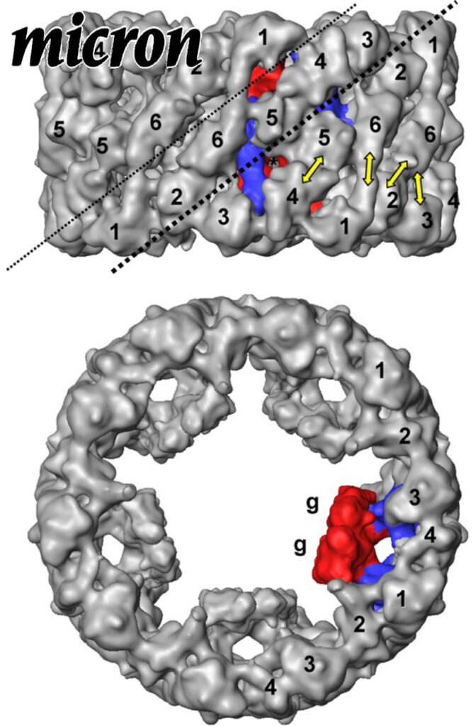
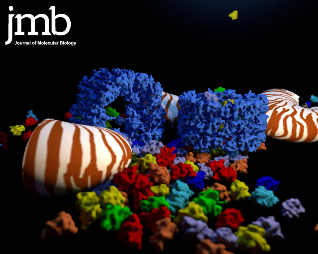
Nautilus pompilius hemocyanin: 9 A cryo-EM structure and molecular model reveal the subunit pathway and the interfaces between the 70 functional units.

Hemocyanins are giant extracellular oxygen carriers in the hemolymph of many molluscs. Nautilus pompilius (Cephalopoda) hemocyanin is a cylindrical decamer of a 350 kDa polypeptide subunit that in turn is a “pearl-chain” of seven different functional units (FU-a to FU-g). Each globular FU has a binuclear copper centre that reversibly binds one O2 molecule, and the 70-FU decamer is a highly allosteric protein. Its primary structure and an 11 Å cryo-electron microscopy (cryo-EM) structure have recently been determined, and the crystal structures of two related FU types are available in the databanks.

Nautilus pompilius hemocyanin: 9 A cryo-EM structure and molecular model reveal the subunit pathway and the interfaces between the 70 functional units. Read More »