Jan-Hannes Schäfer

Acidic Environment Induces Dimerization and Ligand Binding Site Collapse in the Vps10p Domain of Article Acidic Environment Induces Dimerization and Ligand Binding Site Collapse in the Vps10p Domain of Sortilin

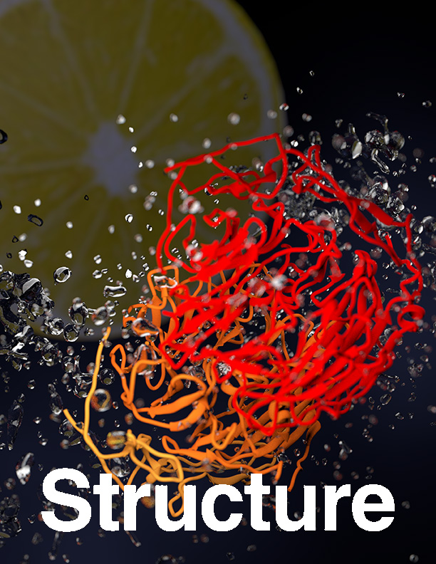
Sortilin is a neuronal receptor involved in transmembrane signaling, endocytosis, and intracellular sorting of proteins. It cycles through a number of cellular compartments where it encounters various acidic conditions. The crystal structure of the sortilin ectodomain has previously been determined at neutral pH. Here, we present the 3.5-Å resolution crystal structure of sortilin at pH 5.5, which represents an environment similar to that of late endosomes, where ligands are released.
Acidic Environment Induces Dimerization and Ligand Binding Site Collapse in the Vps10p Domain of Article Acidic Environment Induces Dimerization and Ligand Binding Site Collapse in the Vps10p Domain of Sortilin Read More »

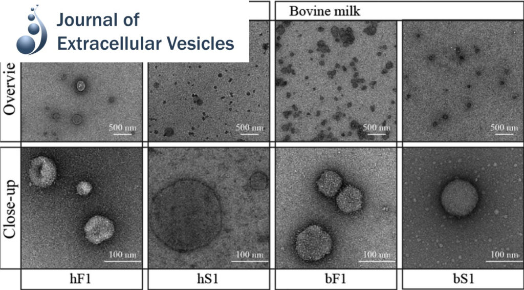
Pellet-free isolation of human and bovine milk extracellular vesicles by size-exclusion chromatography.

Studies have suggested that nanoscale extracellular vesicles (EV) in human and bovine milk carry immune modulatory properties which could provide beneficial health effects to infants. In order to assess the possible health effects of milk EV, it is essential to use isolates of high purity from other more abundant milk structures with well-documented bioactive properties.

Pellet-free isolation of human and bovine milk extracellular vesicles by size-exclusion chromatography. Read More »

Hidden Twins: SorCS neuroreceptors form stable dimers. J Mol Biol

SorCS1, SorCS2 and SorCS3 belong to the Vps10p-domain family of multiligand receptors. Genetic and functional studies have linked SorCS receptors to psychiatric disorders, Alzheimer’s disease and type 2 diabetes, demonstrating critical roles in neuronal functionality and metabolic control.

Hidden Twins: SorCS neuroreceptors form stable dimers. J Mol Biol Read More »

Division of labor in transhydrogenase by alternating proton translocation and hydride transfer.

NADPH/NADP+ (the reduced form of NADP+/nicotinamide adenine dinucleotide phosphate) homeostasis is critical for countering oxidative stress in cells. Nicotinamide nucleotide transhydrogenase (TH), a membrane enzyme present in both bacteria and mitochondria, couples the proton motive force to the generation of NADPH. We present the 2.8 Å crystal structure of the transmembrane proton channel domain of TH from Thermus thermophilus and the 6.9 Å crystal structure of the entire enzyme (holo-TH).

Division of labor in transhydrogenase by alternating proton translocation and hydride transfer. Read More »

Distinct conformational spectrum of homologous multidrug ABC transporters.

ATP-binding cassette (ABC) exporters are ubiquitously found in all kingdoms of life and their members play significant roles in mediating drug pharmacokinetics and multidrug resistance in the clinic. Here, we used single particle electron microscopy (EM) to delineate the entire conformational spectrum of two homologous ABC exporters (bacterial MsbA and mammalian P-glycoprotein) and the influence of nucleotide and substrate binding.

Distinct conformational spectrum of homologous multidrug ABC transporters. Read More »

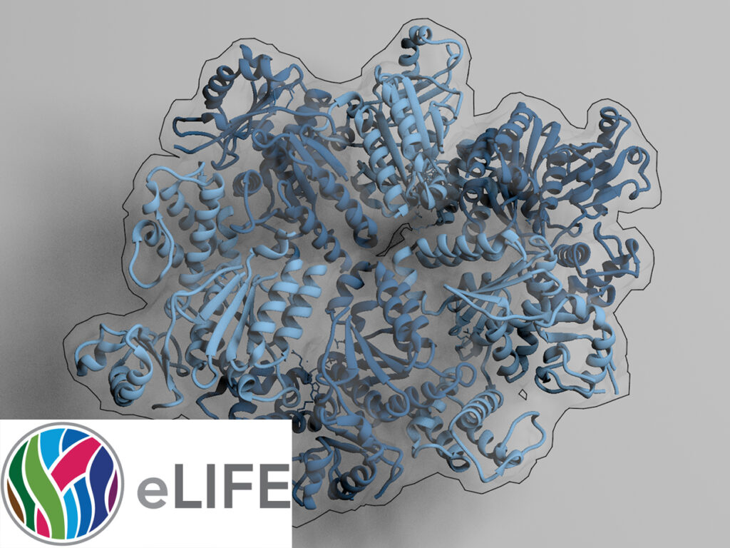
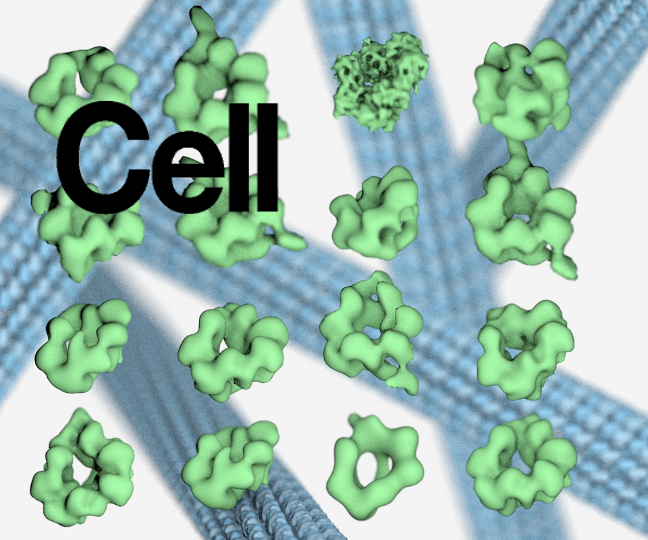
TRIP13 is a protein-remodeling AAA+ ATPase that catalyzes MAD2 conformation switching.

The AAA+ family ATPase TRIP13 is a key regulator of meiotic recombination and the spindle assembly checkpoint, acting on signaling proteins of the conserved HORMA domain family. Here we present the structure of the Caenorhabditis elegans TRIP13 ortholog PCH-2, revealing a new family of AAA+ ATPase protein remodelers.

TRIP13 is a protein-remodeling AAA+ ATPase that catalyzes MAD2 conformation switching. Read More »

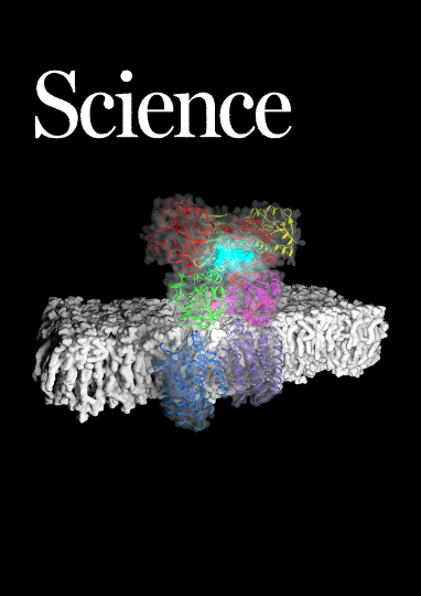
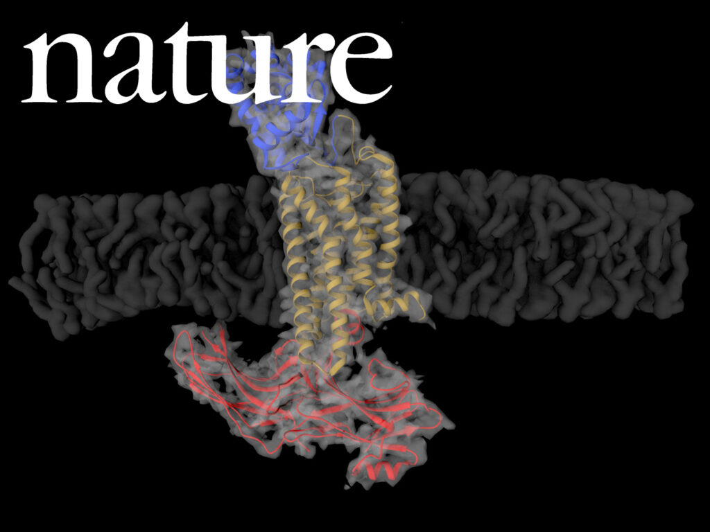
Crystal structure of rhodopsin bound to arrestin by femtosecond X-ray laser.

G-protein-coupled receptors (GPCRs) signal primarily through G proteins or arrestins. Arrestin binding to GPCRs blocks G protein interaction and redirects signalling to numerous G-protein-independent pathways. Here we report the crystal structure of a constitutively active form of human rhodopsin bound to a pre-activated form of the mouse visual arrestin, determined by serial femtosecond X-ray laser crystallography.

Crystal structure of rhodopsin bound to arrestin by femtosecond X-ray laser. Read More »

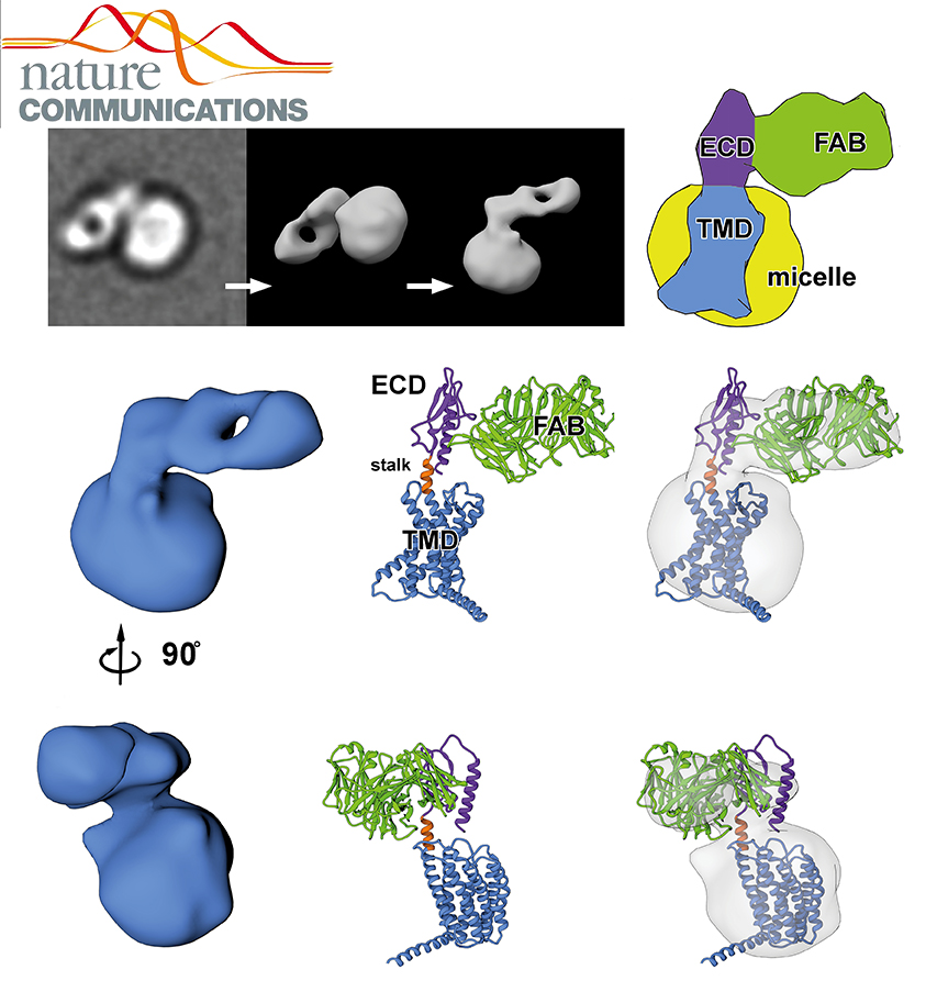
Conformational states of the full-length glucagon receptor.

Class B G protein-coupled receptors are composed of an extracellular domain (ECD) and a seven-transmembrane (7TM) domain, and their signalling is regulated by peptide hormones. Using a hybrid structural biology approach together with the ECD and 7TM domain crystal structures of the glucagon receptor (GCGR), we examine the relationship between full-length receptor conformation and peptide ligand binding.

Conformational states of the full-length glucagon receptor. Read More »

Allosteric communication in the dynein motor domain.

Dyneins power microtubule motility using ring-shaped, AAA-containing motor domains. Here, we report X-ray and electron microscopy (EM) structures of yeast dynein bound to different ATP analogs, which collectively provide insight into the roles of dynein’s two major ATPase sites, AAA1 and AAA3, in the conformational change mechanism.

Allosteric communication in the dynein motor domain. Read More »

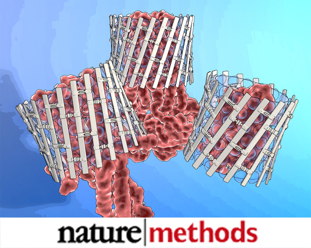
Engineered nanostructured β-sheet peptides protect membrane proteins.

We designed β-strand peptides that stabilize integral membrane proteins (IMPs). β-strand peptides self-assemble in solution as filaments and become restructured upon association with IMPs; resulting IMP–β-strand peptide complexes resisted aggregation when diluted in detergent-free buffer and were visible as stable, single particles with low detergent background in electron micrographs.

Engineered nanostructured β-sheet peptides protect membrane proteins. Read More »